Guadalajara, Jal.

dr.delarosa@uromin.mx
Monday - Friday: 9 am - 8 pm
Saturday: 10 am - 1 pm
Saturday: 10 am - 1 pm

A prostate ultrasound is a simple medical procedure that allows professionals to evaluate the prostate health precisely and quickly. Prostate ultrasound is performed by gently inserting a finger-sized probe into the rectum, allowing for detailed images of the prostate gland and surrounding tissues. This procedure helps doctors detect potential problems or abnormalities quickly and without significant pain for the patient.
Many men feel some concern or curiosity before undergoing this study. It is important to know that preparation for ultrasound is usually minimal, and the results obtained help both in disease detection as in following up on preconditions.
Key Points
- Prostate ultrasound provides an accurate internal view of the gland.
- It is a minimally invasive and quick procedure.
- The results help doctors diagnose and monitor prostate cancer.
What is a prostate ultrasound?
Prostate ultrasound is an imaging test performed to visualize the prostate gland using high-frequency sound waves. This study allows obtaining images in real time and is a key tool in evaluating the size, shape and pathologies of the prostate.
Objective of the procedure
The main purpose of prostate ultrasound is to analyze the prostate in detail without resorting to radiation. Doctors use it to detect changes in the structure of the gland, such as an increase in size (prostatic hyperplasia) or the presence of nodules.
The scan shows possible signs of cancer, inflammation, or infections. In addition, it helps guide procedures such as biopsies when prostate tissue samples need to be taken.
The ultrasound also evaluates the condition of nearby tissues, such as the seminal vesicles. All this is done safely and comfortably for the patient.
Types of prostate ultrasound
Prostate ultrasound can be performed in two main ways: transrectal and Suprapubic. The transrectal technique is the most common and offers sharper images, since the transducer is gently inserted into the rectum to be closer to the gland.
Suprapubic ultrasound, on the other hand, is performed by placing the transducer over the lower abdomen. This option is used in cases where a transrectal approach is not possible or recommended.
Preparing for prostate ultrasound
To obtain accurate images of the size and structure of the prostate, it is essential to take specific steps before the study. Proper preparation can influence patient comfort and the quality of diagnostic results.
Pre-exam recommendations
It is suggested that the patient come with a moderately full bladder, since a full bladder allows a better visualization of the prostate gland during the transabdominal examination. It is usually recommended to drink between 500 ml and 1 liter of water about an hour before the procedure and avoid urinating before the exam.
In the case of transrectal ultrasound, the patient may be instructed to perform an enema the night before or the morning of the study, which helps to clear the rectum and minimize possible image interference. Personal hygiene is important, especially in the perianal area.
Fasting is not commonly required for this study. You should continue with your usual medications, unless otherwise indicated by your doctor. It is helpful to inform the specialist about specific allergies, diseases or treatments before the study.
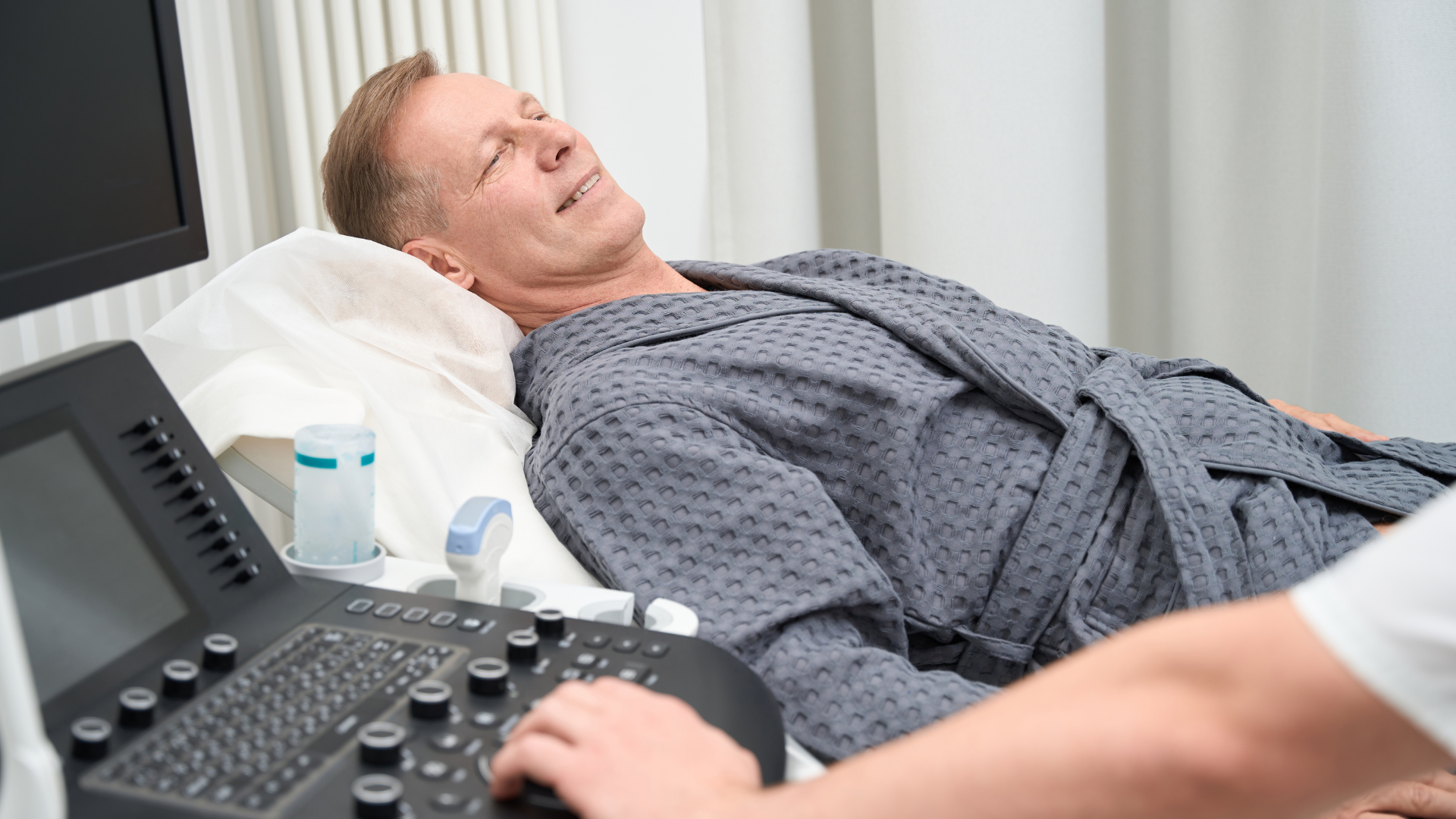
How is prostate ultrasound performed
Prostate ultrasound uses sound waves to generate images of the prostate gland. Depending on the reason, a transrectal or transabdominal technique may be used, both for different purposes and procedures.
Transrectal technique
The transrectal technique is the most common method for accurately visualizing the prostate. It involves carefully inserting a small, lubricated probe into the patient's rectum. This transducer emits sound waves that bounce off the prostate gland, producing detailed images of its different areas.
During the procedure, the patient lies on his side, with his knees slightly bent. The doctor usually uses gel to improve contact and reduce discomfort. The probe collects information in real time, allowing us to observe the size, shape and any abnormality present in the prostate. It is especially useful for guiding biopsies or identifying suspicious nodules.
Transrectal ultrasound may cause mild temporary discomfort, but usually does not require anesthesia. It is a fast and well-tolerated technique, recommended when you need a Accurate diagnosis of the prostate gland.
Transabdominal technique
The transabdominal technique is less used when the objective is to evaluate the prostate in detail, but it is useful in certain cases. It is done by placing a probe over the skin of the abdomen, usually with the patient lying on his back.
To improve image quality, it is recommended that the patient have a full bladder before the examination. Gel is applied to the abdomen to facilitate the sliding of the probe and the transmission of sound waves through the skin. This method is less invasive, since no probe is inserted into the body.
Although it does not offer the same resolution as the transrectal technique, transabdominal ultrasound can detect enlargements, changes in the contour and volume of the prostate. It is useful as a first approach or in patients who do not tolerate the transrectal route well.
Duration and basic steps
The total exam time is usually between 15 and 30 minutes. The process includes several essential steps to ensure clear and reliable images.
- Preparation: The patient should remove clothing from the waist down (transrectal) or make sure their bladder is full (transabdominal).
- Positioning: Depending on the technique, the patient lies on the examination table, on his side or on his back.
- Gel application: Gel is placed on the probe or directly on the skin.
- Insertion or support of the probe: In the transrectal route, the probe is gently inserted into the rectum; in the transabdominal route, it slides over the abdomen.
- Capturing images: The professional moves the catheter slowly to get different views of the prostate.
- Completion: The probe is removed, the gel is cleaned and the patient can be dressed again immediately.
The results are usually ready in a short time and there is no need for special recovery. The person can return to their normal routine after the procedure. Each step is designed to maximize the safety and diagnostic quality of the study.
Interpretation of outcomes and aftercare
After performing a prostate ultrasound, it is essential to understand both the normal sensations after the procedure and what the results mean and when to seek additional medical attention. Proper care and follow-up facilitate an uncomplicated recovery and a correct interpretation of the findings.
What to Expect After the Procedure
It is common for the patient to experience mild rectal discomfort after the examination if it was performed transrectally. You may feel a slight irritation or urge to poop for a few hours afterward. In rare cases, a small amount of bleeding may be seen in the stool, urine, or semen, which usually goes away in a short time.
In most cases, normal activity can be resumed right away. Driving is not recommended if sedatives were administered, but this is rarely necessary for routine prostate ultrasound. If symptoms such as severe pain, fever, or heavy bleeding occur, it is important to see a doctor right away.
Meaning of the findings
The results of prostate ultrasound report key characteristics of the gland, such as size, shape, presence of suspicious nodules or lesions, and alterations in structure. These findings help detect diseases such as benign prostatic hyperplasia, infections, or tumors.
A report may include tables with prostate measurements and detailed descriptions of any anomalies detected. If biopsies are taken during the procedure, the anatomopathological results will be available later and will be essential to confirm or rule out the presence of prostate cancer.
Follow-up inquiries
Depending on the results, the health professional may recommend regular checkups, additional tests such as MRI or biopsy, or simply clinical observation if the findings are not relevant. It is important for the patient to keep a record of symptoms and share them at future appointments.
Any medication instructions or restrictions suggested by the doctor should be followed to the letter. A follow-up schedule makes it possible to schedule future ultrasound or related tests, thus ensuring adequate control of prostate health. If doubts or new symptoms arise, it is advisable to contact the specialist without delay.
Frequently Asked Questions
Prostate ultrasound is a commonly used test to evaluate the size, shape and possible abnormalities of the prostate gland. There are different ways to do this, and the preparation, the process and its usefulness may vary depending on the method used.
How do you prepare for a prostate ultrasound?
Preparation depends on the type of ultrasound. For a transrectal ultrasound, an enema is usually recommended before the exam to clean the rectum, helping to obtain clearer images.
In the case of suprapubic or transabdominal ultrasound, the patient may need to drink water to keep the bladder full. This makes it easier to see the prostate from the abdomen.
Is the prostate ultrasound procedure painful?
A prostate ultrasound usually doesn't cause significant pain. The transrectal procedure can be uncomfortable, as the transducer is inserted into the rectum, but the discomfort is usually mild and momentary.
During the examination, the patient may experience pressure, but he should not feel severe pain.
Can ultrasound detect prostate cancer?
Prostate ultrasound helps identify suspicious areas and abnormalities in the gland. However, this method cannot confirm prostate cancer on its own.
Ultrasound can guide a biopsy when irregular areas are detected or nodules in the prostate.
What size of the prostate is considered normal on an ultrasound?
Normal prostate volume is usually between 20 and 30 ml in adult men. However, the size may vary depending on age and individual factors. An increase in prostate size may be associated with benign hyperplasia or, in some cases, with other pathologies.
What protocol is followed in a transabdominal prostate ultrasound?
On a transabdominal ultrasound, the patient must usually come with a full bladder. You are asked to lie in a supine position while a gel is applied to your lower abdomen. The technician moves the transducer over the suprapubic area to obtain images of the prostate.
Preparing yourself well is the first step towards an accurate diagnosis
Understanding how a prostate ultrasound is performed and what preparation it requires allows you to face the study with greater peace of mind and confidence. This fast and minimally invasive procedure provides essential information for evaluating prostate health and guiding sound clinical decisions.
In Precision Prostate Clinic, each evaluation is carried out with precision, respect and state-of-the-art technology. Our team is committed with offering a safe and professional environment from the first contact.
Tag:
NEWSLETTER
Subscribe our Newsletter
To get latest updates, News, Technology we use and offers
Thank you! Your submission has been received!
Oops! Something went wrong while submitting the form.
