Guadalajara, Jal.

dr.delarosa@uromin.mx
Monday - Friday: 9 am - 8 pm
Saturday: 10 am - 1 pm
Saturday: 10 am - 1 pm
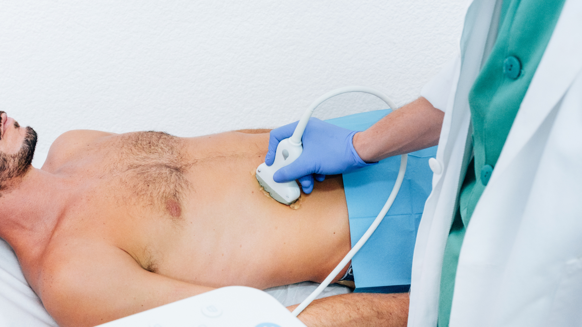

Transabdominal ultrasound for prostate is a non-invasive technique used to visualize and evaluate the size and structure of the prostate gland in men. This method makes it possible to measure prostate volume and detect anatomical changes quickly and comfortably for the patient.. It is usually done with the patient having a moderately full bladder, which improves the quality of the image obtained during the study.
During the procedure, a transducer is used on the lower abdomen with frequencies of 2 to 5 MHz, providing standard images and avoiding discomfort associated with other more invasive techniques. It is an option used for monitoring urinary symptoms, evaluating prostate size and in settings where other modalities are not indicated or are not tolerated.
Key points
- It allows a quick and convenient evaluation of prostate size.
- It is a versatile tool that is well tolerated in a variety of clinical indications.
- It has limitations for the diagnosis of prostatic neoplasms.
Fundamentals of transabdominal ultrasound for prostate
Transabdominal ultrasound is a useful imaging technique for prostate evaluation, especially in settings where access to transrectal ultrasound is limited. This method makes it possible to observe the size and structure of the prostate without requiring a direct invasion of the rectum.
Principle and operation of transabdominal ultrasound
Transabdominal ultrasound for the prostate uses high-frequency sound waves, usually between 2 and 5 MHz. The transducer is placed over the lower abdomen with conductive gel to ensure good wave transmission.
These waves pass through the full bladder and reach the prostate, generating images in real time. It is essential for the patient to have a moderately full bladder to displace the intestine and improve visualization.
This method is especially useful for determining prostate volume and detecting enlargement or structural changes. However, their ability to identify small lesions or distinguish tumors is more limited than that of other techniques, such as transrectal ultrasound.
Differences between transabdominal and transrectal ultrasound
Transabdominal ultrasound is less invasive and more comfortable for most patients. This exam is performed on the skin of the abdomen, avoiding the discomfort of the rectal approach.
In contrast, transrectal ultrasound provides greater anatomical detail and can better detect small or suspicious lesions in the prostate. For this reason, transrectal surgery is often preferred for the detection and monitoring of prostate cancer.
The following table compares both methods:

Patient preparation
Before the examination, the patient must present himself with a moderately full bladder. This improves visualization of the prostate, as an empty bladder can make it difficult to obtain clear images.
It is recommended to drink 500 to 1000 ml of water at least one hour before the procedure. Fasting or stopping regular medications is not needed for this test.
The patient should wear comfortable clothing and may need to remove clothing from the waist down. Some clinics recommend attending accompanied for reasons of comfort and mobility.
Technique of realization
During the examination, the patient lies on the stretcher in a supine position. The professional applies conductive gel to the lower abdomen to facilitate contact of the transducer with the skin.
The transducer glides smoothly over the suprapubic area. The operator adjusts the position and angle to obtain transverse and longitudinal images of the prostate and nearby structures, such as the bladder and seminal vesicles.
The technique uses linear, convex or sector transducers with frequencies between 2 and 5 MHz, as detailed in this prostate ultrasound study. The process usually takes 10 to 20 minutes and is painless.
Prostate volume measurement
To measure prostate volume, images are obtained in the sagittal and transverse planes. Three key dimensions are identified: width (lateral), height (anteroposterior) and length (cranio-caudal). This method provides an accurate estimate of prostate size in clinical and research routines.
The measurement makes it possible to compare against reference values for age, to aid in the diagnosis of benign prostatic hyperplasia and to decide on the need for additional treatments.
Clinical Indications and Main Applications
Transabdominal ultrasound is a non-invasive tool that provides relevant anatomical and functional information about the prostate gland. It allows for the rapid and safe evaluation of various urological conditions affecting adult men.
Benign prostatic hyperplasia detection
La benign prostatic hyperplasia (BPH) is a common cause of prostate enlargement in older men. Transabdominal ultrasound allows precise measurement of prostate volume, which is essential for clinical decision-making.
This method helps differentiate benign growth from other pathologies that can also produce obstructive urinary symptoms. It is possible to identify changes in the architecture of the prostate and to delimit areas affected by hyperplasia.
In some cases, invasive procedures can be avoided when prostate volume does not justify major interventions, as indicated in the evaluation of prostate size before BPH surgery.
Frequently Asked Questions
Transabdominal prostate ultrasound makes it possible to evaluate the prostate gland and detect enlargements, abnormal formations or alterations in its morphology. Differences from other methods and clinical indications depend on the patient's context and the objective of the study.
What is the standard protocol for performing a transabdominal ultrasound of the prostate?
Generally, the patient should come with a moderately full bladder, as this improves prostate visualization through the abdominal wall. You are asked to drink water before the exam and avoid evacuating your bladder until the procedure is complete.
The scanner places the transducer in the lower abdomen and uses conductive gel to optimize the transmission of ultrasound waves. Images are obtained in longitudinal and transverse planes to fully study the gland.
In which cases is it preferable to use transabdominal ultrasound rather than transrectal ultrasound to evaluate the prostate?
Transabdominal ultrasound is preferred when there are contraindications or rejections for the transrectal approach, such as anal fissures, infections, active hemorrhoids, or a history of rectal surgery. It is also useful for initial controls and evaluation of prostate volume in settings where malignancy is less suspected.
In pediatric or young patients, where tolerance to a rectal approach may be lower, abdominal access is less invasive.
What are the key anatomical findings in a prostate ultrasound?
The contour of the prostate, its echogenicity (homogeneous under normal conditions) and the overall size are observed. The periurethral tissue, the prostate capsule and the peripheral and central areas can be distinguished in good quality images.
Alterations such as nodules, calcifications or hypoechoic areas may indicate benign or malignant pathologies.
What measures are crucial during prostate ultrasound and how are they interpreted?
The anteroposterior, transverse and longitudinal diameter of the prostate makes it possible to calculate its total volume, essential for the diagnosis of benign hyperplasia and for planning treatments or surgeries.
A volume greater than 30-40 cc is usually associated with obstructive symptoms. In addition, possible injuries, suspicious foci and the thickness of the residual tissue after urination are evaluated.
How is transabdominal ultrasound different from other imaging techniques for the detection of prostate diseases?
Transabdominal ultrasound is a fast, accessible and non-invasive technique, but it has a lower resolution for detecting small lesions compared to transrectal ultrasound or magnetic resonance imaging.
It does not use radiation and is especially valuable for initial evaluations or in patients who are limited to other tests. It allows general anatomical assessments and the monitoring of prostate size.
Which prostate pathologies are most adequately diagnosed using transabdominal ultrasound?
Benign prostate hyperplasia and large cystic formations or abscesses are easily identifiable with this technique. The method is less sensitive for detecting small tumors or lesions suspected of cancer, and these are better evaluated by transrectal ultrasound or resonance imaging.
It is also useful for detecting urinary retention, prostate stones and evaluating the relationship of the prostate with the bladder.
A Key Tool for Prostate Care
Transabdominal ultrasound is an accessible, safe and effective technique for obtaining an overview of the state of the prostate. Although it is not a substitute for more detailed studies such as transrectal ultrasound, it plays an important role in the initial diagnosis and monitoring of certain urological conditions.
In Precision Prostate Clinic, we integrate this type of study into a complete and personalized diagnostic approach, supported by advanced technology and a highly specialized medical equipment.
Tag:
NEWSLETTER
Subscribe our Newsletter
To get latest updates, News, Technology we use and offers
Thank you! Your submission has been received!
Oops! Something went wrong while submitting the form.
